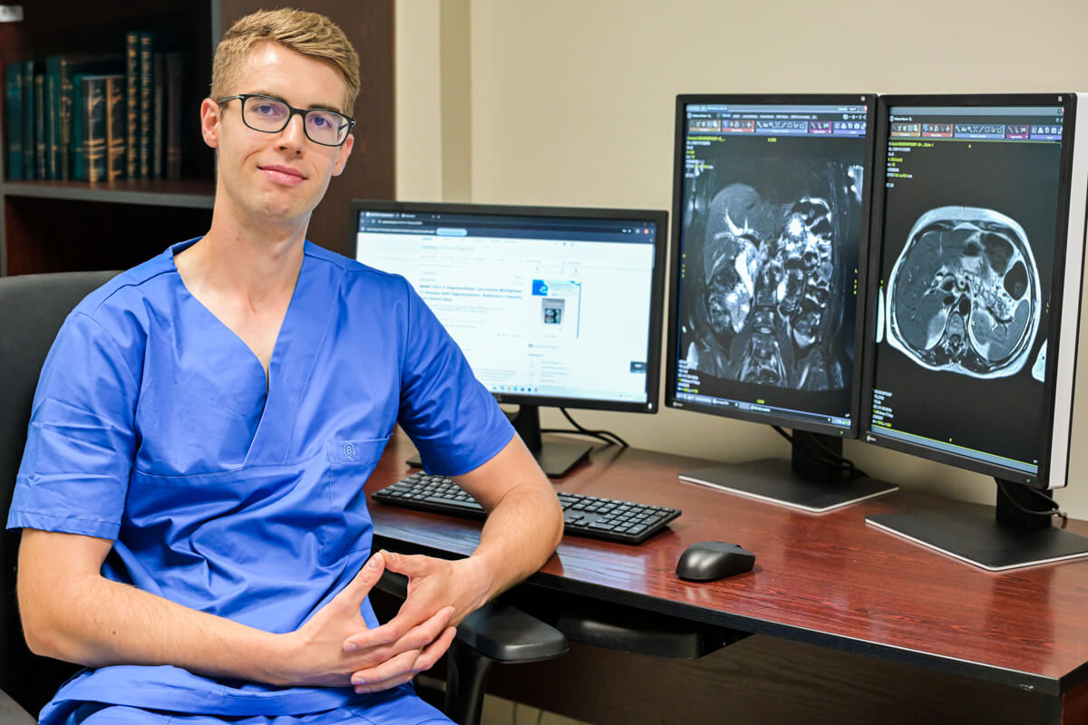
The research focuses on applying machine learning and deep learning methods to analyze medical images, primarily computed tomography (CT) and magnetic resonance imaging (MRI). Why? Because in the diagnosis and treatment of cancers, imaging studies are often the first to signal a problem and accompany the patient throughout all stages of care.
“The development of modern imaging techniques, such as CT and MRI, provides significantly richer diagnostic data than what radiologists traditionally use. In other words, large amounts of data are available but are not always fully utilized – in clinical practice, usually only part of the information visible in the images is analyzed. Meanwhile, clues that can help predict disease progression and treatment response are hidden in the structure, shape, vascularization, and dynamics of tumor changes. This means that advanced image analysis – for example, one using artificial intelligence – can open a new chapter in personalized oncology,” writes Dr. Krzysztof Bartnik in his scientific article.
Readers are encouraged to read the full article, published in the Science!
Link to the page with the article “Artificial intelligence as a step toward personalized oncology”