Artificial intelligence – a step towards personalized oncology
The project titled “Artificial intelligence in predicting treatment outcomes for abdominal cancers: implementing machine learning and deep learning methods for the comprehensive analysis of medical imaging”, run by Krzysztof Bartnik, MD, PhD, and carried out in consortium with the Warsaw University of Technology and Duke University (U.S.), has received funding from the National Science Centre under the SONATINA grant program.
Neoplasms – one of the biggest health challenges
One in five people across the world will develop some kind of cancer in their lifetimes. In Poland, 170,000 people develop it every year. The disease poses an immense challenge not just to patients, but also for the healthcare system, which has to detect it as soon as possible and plan effective treatment. Radiology is crucial in both diagnostics and therapy: imaging tests such as tomography and MRI are often the first to pick up the issue, and they are subsequently involved at each stage of treatment.
As the most common type of primary liver cancer and one of the most dangerous abdominal cancers, hepatocellular carcinoma is a special type of neoplasm. It is complicated to treat, and predicting treatment outcomes remains a huge challenge. Every step towards improved predictions, more accurate treatment planning, and more personalized therapy makes a real difference in the lives of thousands of patients.
An effective fight against cancer starts with an accurate prognosis of the course of the disease
Traditional forecasting methods, based mostly on the simple characteristics of the tumor such as its size and growth dynamics, have proven to be insufficient. That is because they often do not reflect the actual uniqueness of the patient and their condition. For two patients with tumors of similar size, the course of the disease may be totally different, which is due to factors such as liver condition, comorbidities (if any), the biological subtype of the cancer, or response to previous lines of treatment. This means that forecasting the course of the disease and treatment efficacy require a much more complex approach.
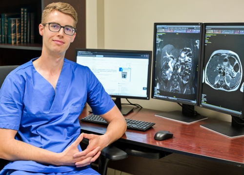
Modern radiology techniques in the fight against cancer
The development of modern imaging techniques such as computed tomography (CT) or magnetic resonance (MRI) has offered much richer diagnostic data than those used historically by radiologists. In other words, large amounts of data are available, but not all of them are used to the full extent – in clinical practice, only some of the information visible in the images gets analyzed. Yet tumor structure, shape, vascularization or growth dynamic offer hints that may help predict the course of the disease and response to treatment. This means that advanced image analysis – with the use of artificial intelligence, for instance – may open a new chapter in personalized oncology.
Modern imaging goes beyond just ‘photographing the patient’. Each CT or MRI scan generates hundreds of cross-sections and millions of pixels, each of which carries information about the structure, density, vascularization, or tissue function. Those images may offer tens or even hundreds of features describing the tumor: from simple geometrical parameters to complex texture indicators or contrast dynamics.
This creates vast, multi-dimensional imaging datasets that defy the computing capacity typically used with traditional statistical methods. Working on such datasets requires tools capable of capturing subtle patterns that escape the human eye. This is where artificial intelligence (AI) may step in. Our project is aimed at developing a new class of AI models that will allow for predicting long-term treatment outcomes based on the advanced analysis of CT and MRI images. The models will look not only at the tumor itself, but also abdominal organs such as the liver or spleen.
Why is AI a game changer in forecasting?
Artificial intelligence algorithms used so far in oncology have mostly focused on analyzing the tumor itself – its volume or shape. Yet the patient’s clinical condition also depends on the functioning of nearby organs and the presence of comorbidities. The diseases that affect internal organs often significantly alter their morphology in imaging tests, e.g. by changing the structure of the organ – which is not a standard aspect covered in image descriptions. By extending the analysis to include the characteristics of multiple organs visualized in the image, we will be able to capture that complexity and significantly improve the accuracy of predictions.
Our project combines two approaches:
- deep learning, which allows computers to ‘learn’ how to analyze data by recognizing patterns and dependencies. In our case, this will allow for quickly and automatically segmenting multiple areas of interest (e.g. tumors or internal organs) pictured in medical images;
- machine learning. This is a broader category of analytical methods, which – based on pre-defined properties extracted from the images – build prognostic models. Unlike deep learning, where the computer itself extracts features from data, classic machine learning is based on parameters defined by experts (e.g. tumor shape, texture, contrast dynamics, etc.). This allows for predicting patient life expectancy or progression-free survival.
By combining these two approaches, we will be able to reduce the need for manually segmenting radiological images, which is a time-consuming process, and then to extract calculation variables from them. For years, these limitations have been one of the main challenges in radiomics, a field concerned with the extraction of a large number of hidden features from medical images and analyzing them in order to better understand the biology of tumors and predicting the course of the disease.
Artificial intelligence meets radiology: foundation models
At this point, it is worth mentioning that the project uses foundation models, or advanced artificial intelligence algorithms trained on vast, diversified sets of data. Their capacity to ‘learn’ universal representations allows for adjusting them easily for different tasks – from segmentation to predicting treatment outcomes. In radiology, this opens up an opportunity for improved analysis of rare clinical cases, or for more accurate diagnostics.
During his recent visit at the WUM, Professor Maciej Mazurowski, Head of the Duke Center for Artificial Intelligence in Radiology, discussed projects such as The Human Body Project, aimed at creating a comprehensive “foundation model” for the entire human body. The model will largely support the solutions tested as part of our project.
More information about Professor Mazurowski’s visit and lecture at WUM can be found here
A project that will improve the understanding and treatment of liver cancer
Our project will bring a number of benefits. Firstly, for patients and doctors, as it will improve the quality of predictions for the course of the disease and treatment efficacy while reducing the burden on radiologists thanks to the automation of analysis.
Secondly, it involves developing open datasets for the scientific community. Finally, it will enhance international and cross-disciplinary cooperation – between doctors, IT specialists, and biologists.
Most importantly, the current project is an excellent prelude to subsequent research initiatives.
What will be done as part of the project?
The project will include three main stages:
- Improving computed tomography models for hepatocellular carcinoma (HCC)
Based on the clinical datasets held by the WUM UCC, prediction models based on multi-organ segmentation will be developed. New imaging predictors will be added to the set of radiomic features already in use. - Applying the methodology to another abdominal cancer
In order to verify how universal this approach may be, the models will be tested in an alternative oncological scenario. This will be done using federated learning, a modern method of distributed learning that allows for training algorithms on data from various centers without the necessity to transmit them. This is crucial with respect to the protection of patient data and research scalability. This stage will be done in partnership with Duke University (U.S.), which is a global leader in AI in radiology. - Creating a unique MRI dataset for patients with hepatocellular carcinoma
With the high resolution of tissue imaging, magnetic resonance is the golden standard in primary liver cancer diagnostics. However, the collections of magnetic resonance imaging and images of hepatocellular carcinoma available publicly are extremely limited. Thanks to the resources from one of the largest hepatology centers in Europe, we will create a dataset based on a standardized imaging protocol. Making such a dataset available will open the door to global research into the use of AI for hepatocellular carcinoma.
The power of cooperation between experts in medicine and new technologies
The SONATINA-funded project will be carried out in cooperation with Duke University and the newly created Centre for Credible AI (CCAI) at the Warsaw University of Technology. The Centre is a university-wide unit that develops and implements AI solutions that guarantee safety, transparency, and reliability.
The CCAI operates at the junction of information technology, mathematics, and medicine, and has ambitious goals: building AI systems that can be understood, verified, and monitored, developing standards, and supporting the education of a new generation of artificial intelligence experts.
The synergy between radiology research at the WUM, the experience of Duke University, and CCAI infrastructure opens the door for projects with an even greater reach, in both scientific and clinical terms. The team has already been working on new grant applications, aimed at boosting Poland’s position on the global map of research into the medical applications of AI.
But apart from the Warsaw University of Technology and Duke University, we must also highlight the importance of teamwork. Such projects would not be possible without the commitment of radiologists, technicians, and nurses from the 2nd Department of Radiology WUM. The inherent cooperation with surgeons, neurosurgeons, geneticists, pathologists, oncologists, and many other specialists is of utmost importance, too. Success is only possible if we work as a team.