How to tell what’s going on in the circulatory system just by looking at your eyes

Maria Żmijewska is the author of the study titled “Retinal Microcirculation Measurements in Response to Endurance Exercises Analysed by Adaptive Optics Retinal Camera,” which investigated the effect of physical exercise on retinal microcirculation.
Why is this important?
Retinal microcirculation has long attracted the interest of physicians across various specialties. The eye is the only organ in which the smallest arterioles can be visualized noninvasively, and their responses to different factors can be analyzed. Moreover, retinal vessels are of the same type as coronary and cerebral vessels, meaning that anatomically they respond in the same way as all end vessels. A key mechanism that enables the maintenance of stable microcirculation in three vital organs – the heart, brain, and eye – regardless of fluctuations in blood pressure, is the myogenic self-regulatory system. If this mechanism can be assessed noninvasively by examining the eye, then the analysis of retinal vessels becomes a reliable and increasingly used marker for evaluating cardiovascular risk.
Exercise, blood vessels, and myogenic self-regulation
Physical exercise is beneficial to health because it improves blood oxygenation and increases cardiac performance. However, the exercise-induced increase in blood pressure and blood flow must be controlled by a properly functioning myogenic self-regulatory mechanism. Otherwise, vessel rupture could occur.
Myogenic self-regulation occurs primarily in small blood vessels, arterioles, and precapillary sphincters. How does this mechanism work? In response to increased arterial pressure and perfusion pressure, defined as the difference between arterial inflow pressure and venous or tissue outflow pressure in a given organ, the precapillary sphincter smooth muscle contracts. This leads to an increase in peripheral resistance and, consequently, a reduction in local blood flow.
The phenomenon of self-regulation resulting from vessel contraction was first described in 1902 by Bayliss, although only in postmortem histological preparations.
Myogenic self-regulation decreases with age. Passive stretching of arterioles induces their contraction and closure of sphincters, resulting in cessation of blood flow, while reduced stretching produces the opposite effect, leading to dilation and opening of precapillary sphincters with subsequent hyperemia. As self-regulation declines, vessels lose the ability to increase resistance in response to exercise. This increases the risk of retinal and cerebral hemorrhages in older individuals, not only during physical exertion but also during fluctuations in blood pressure.
The use of “space-age” imaging
In this study, I used the RTX 1 microscope located in the Chair and Department of Ophthalmology at WUM’s Faculty of Medicine, at the Independent Public Clinical Ophthalmology Hospital on Sierakowskiego Street, headed by Prof. Jacek P. Szaflik. The RTX 1 microscope is based on adaptive optics technology, which provides image resolution comparable to histological resolution.
Adaptive optics was first introduced by astronomer Horace Babcock in 1953. This technique made it possible to minimize “turbulence” caused by atmospheric vibrations, which make stars appear to flicker to the naked eye, and to obtain much clearer images in astronomical telescopes.
The use of such a highly sensitive microscope enabled me to visualize photoreceptors and the smallest retinal arteries, including those measuring only several dozen micrometers. Adaptive optics technology is also a very effective tool for assessing the risk of cardiovascular events and monitoring their progression. The results made it possible to analyze changes in vessel lumen diameter and wall thickness in response to submaximal aerobic exercise that met the criteria of a cardiac stress test.
What was measured in the study?
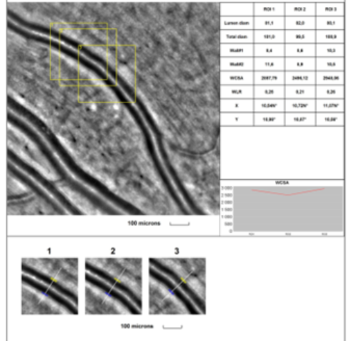
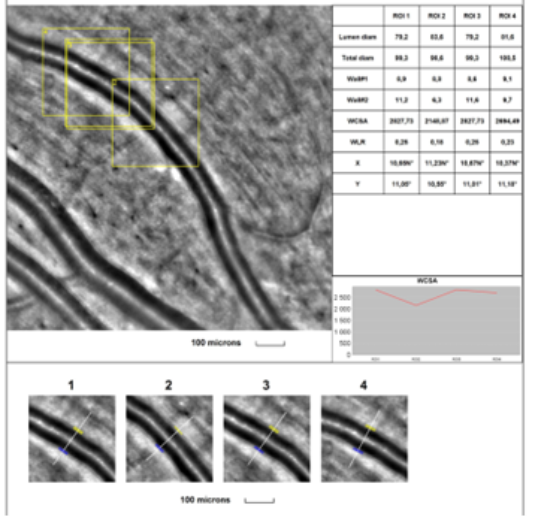
The same retinal arterial vessel, with a diameter of 70 to 130 μm, was measured before and immediately after exercise, within the first two minutes. The study was conducted in healthy volunteers, approximately 20 years old, who exercised on a rowing ergometer until reaching a submaximal heart rate. The study was supervised by an ophthalmologist and a cardiologist.
What was demonstrated? That intense physical exercise, accompanied by an increase in systolic blood pressure and heart rate, leads to constriction of small retinal arteries. A decrease in vessel diameter and wall thickness was observed, along with a significant reduction in the wall-to-lumen ratio (WLR) and wall cross-sectional area (WCSA).
Before:
After:
A major step in studying self-regulation
My study is the first noninvasive investigation to confirm retinal vessel constriction during exercise and the presence of physiological myogenic self-regulation mechanisms in retinal microcirculation. Previously, this phenomenon was documented by Harris A. (Harris A et al. Retinal blood flow during dynamic exercise. Graefes Arch Clin Exp Ophthalmol. 1996), but that study used invasive fluorescein angiography.
The use of adaptive optics to study retinal arteries offers an opportunity for early detection of abnormalities in myogenic self-regulation in patients who cannot undergo invasive procedures. It also allows visualization of early arterial changes in hypertension, which in the near future may become an essential component of diagnostic evaluation in most, if not all, patients with hypertension.