Extended right hemihepatectomy is currently the most extensive, feasible liver resection surgery.
The standard for performing such large resections is laparotomy - a wide opening of the abdominal cavity under the costal margin. This extensive access provides an adequate view of the surgical field and facilitates the liver resection, but it also has disadvantages for the patient: cutting into the abdominal wall exposes the patient to greater postoperative pain and other complications associated with healing of an extensive surgical wound. It also prolongs recovery in the postoperative period. This is important because shortening the rehabilitation period is crucial for early initiation of complementary chemotherapy.
An additional benefit of laparoscopic surgery is more precise tissue dissection, which reduces intraoperative blood loss and minimizes the risk of blood transfusion.
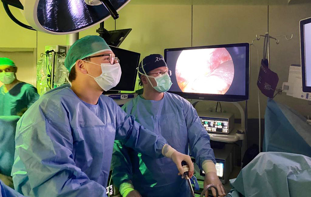
The use of laparoscopy in large liver resections is limited by the considerable difficulty of the procedure - therefore only a few centers in the world perform it.
Performing the most extensive of these surgeries was possible thanks to the experience that Professor Michał Grąt and his team gained while creating and developing the pioneering program of extensive laparoscopic liver resections in the Department of General, Transplant and Liver Surgery, headed by Professor Krzysztof Zieniewicz.
Extremely important for the performed procedure was the early occlusion of the venous blood inflow and outflow from the right liver segments, which led to hypertrophy of the healthy part of the liver that took over the function of the whole organ after the resection. A team of interventional radiologists led by Professor Olgierd Rowinski of the Second Department of Clinical Radiology was responsible for this part of the preparation for the surgery. Through these coordinated clinical efforts, a patient with an initial inoperable advanced biliary tract cancer received treatment that enabled rapid recovery through the use of modern, minimally invasive procedures.
